Despite their initial description 180 years ago, scientists for many years failed to understand the significance of the lone hair-like structures that extend from the surface of many cell types, including epithelial cells and neurons.1 Known as primary cilia, the physical differences between these immotile organelles and fluid-propelling motile cilia were not apparent. In the 1950s, transmission electron microscopy enabled researchers to distinguish the ultrastructure of these two forms of cilia and how primary cilia function in human health and disease.2
Now, in a recently published Journal of Cell Biology paper, scientists used a newer electron microscopy technique, called volume electron microscopy (vEM), to examine how primary cilia on developing neurons change during differentiation.3 The research provides valuable insights into a process critical for developing and adult tissues of the brain and beyond.
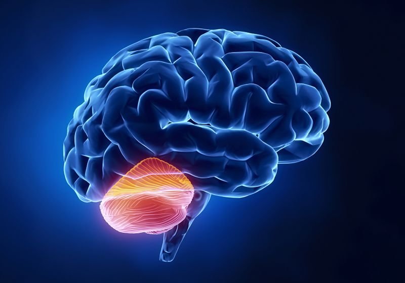
Carolyn Ott and her team discovered how differentiating granule cells in the cerebellum permanently deconstruct their primary cilia.
Matt Staley
Primary cilia are important sensory organelles that play a role in cell signaling and development. “Sometimes people think of [primary cilium] as an antenna because they are coated in receptors and the kinds of messages cilia can receive depend on what receptors the cell expresses and moves to the cilia,” said Carolyn Ott, a cell biologist at Howard Hughes Medical Institute and coauthor of the study. “So, it is a tunable signaling structure.”
Although numerous processes within the brain involve primary cilia, researchers understand very little about how modifications to the organelle’s ultrastructure affect neurodevelopment. Immature granule cells (GCs), which become the most abundant neuron type within the adult brain, offer a unique opportunity to answer this question.
During development, the ligand sonic hedgehog (SHH) binds to receptors on the primary cilia of progenitor GCs, leading to the cells’ proliferation, differentiation, and migration to deeper layers of the cerebellum.4 But, once the differentiation stage begins, the cells stop responding to SHH. Additionally, immunofluorescence analysis has revealed that differentiating GCs have fewer and shorter cilia than their immature counterparts, with adult GCs rarely possessing these structures. This suggests that these cells disassemble their cilia during maturation. However, examining enough of these organelles to fully characterize this disassembly process presented a challenge for the researchers.
“Cilia are hard to find if you are doing conventional EM,” said Ott. “They are low-frequency structures. There is one cilium per cell.”
To analyze more primary cilia, Ott and her team examined publicly available vEM datasets of developing mouse cerebella. Because the GCs’ developmental stage corresponds to their depth in the tissue, a single reconstructed volume captures cells from different phases of maturation. From these volumes, Ott and her team determined that primary cilia permanently disassemble as GC progenitors differentiate. Given that this disassembly occurs in postmitotic cells, the researchers named this process cilia deconstruction to distinguish it from the transient cilia disassembly that occurs prior to cell division.

Through volume electron microscopy, the researchers observed that differentiating granule cells frequently conceal their disassembling primary cilia (cyan) from the extracellular environment. Primary cilia originate from mother centrioles (purple), which dock at the plasma membrane during this deconstruction process.
Carolyn Ott
While examining the ultrastructure of the primary cilia during cilia deconstruction, the researchers observed that many differentiating GCs possessed short cilia that were completely enclosed within a membranous compartment in their cytoplasm. This concealed these disassembling structures from stimuli in the extracellular environment, such as SHH.
During cilia deconstruction, Ott and her team also noted that mother centrioles, which are responsible for the initial formation of cilia, ultimately dock at the plasma membrane but remain unciliated. This surprised the researchers as scientists have only observed a few examples of centrioles docking without forming cilium. The researchers detected novel deconstruction intermediates that were different from those observed during premitotic cilia disassembly, suggesting distinct processes. However, the team was unable to order the intermediates into a single linear pathway. Instead, they suggested that there are several cilia deconstruction routes possible that could generate unciliated adult GCs with docked mother centrioles.
“This is a nice study that combines some new technology and analysis to answer some questions about primary cilia that had not been well studied before and comes up with some interesting and unexpected findings that are going to be important for the field and the broader cell biological community,” said David Breslow, a cell biologist at Yale University who was not involved in the study. “An interesting question that emerges is how many different ways to disassemble cilia are there and what versions of these processes are being used in different physiological contexts.” Breslow hopes that researchers will realize the potential of re-analyzing these information-rich vEM datasets to gain new insights into the brain and other tissues. “Now we are realizing, through works like this, that these datasets can be mined for other types of information relevant to cilia or other cell biological phenomena.”
While cilia deconstruction could be important for the differentiation of other ciliated progenitors, this process may also play a role in medulloblastoma development. These brain tumors result from aberrant GC proliferation in the cerebellum, and in some types of this cancer, tumor cells possess primary cilia and respond to SHH signaling.5 This suggests that hiding and breaking down cilia is important to prevent continuous SHH stimulation, which could cause cancer. “Understanding what is happening to cilia may be important to understanding what is gone wrong in the tumor, which will hopefully lead to new targets,” Ott said.


