Magdalena (Magda) Zernicka-Goetz, today a developmental and stem cell biologist at the University of Cambridge and California Institute of Technology, recalled being an artistic child who enjoyed creating things with her hands. In high school, she planned to study neuroscience because she was fascinated by how the brain worked, but when she saw an embryo for the first time, in her first year of college, it called to her aesthetic sense. “When you see them developing, you are also taken by the mystery of the process and magic of that process,” Zernicka-Goetz explained. “I was interested by the beauty, but not just this beauty on the surface, but also this beauty deep down there. How does this work?”
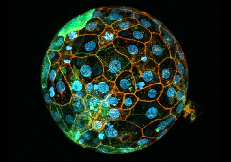
Magdalena Zernicka-Goetz studies human and mouse embryos to learn how the cells mature and form complex structure.
The Gladstone Institute
This chance exposure to the world of developmental biology set Zernicka-Goetz on a course to explore the secrets of the embryo. She received her PhD at the University of Warsaw under Andrzej Tarkowski and completed two postdoctoral fellowships before starting her own lab at the University of Cambridge in 1997. In 2019, she started a second lab group at the California Institute of Technology. In both labs, she studies how stem cells differentiate and develop in the embryo.

“I look [at] how cells become different from each other in scientific journey,” Zernicka-Goetz said about her work. “I embrace those differences; however small and big they are because even small differences can lead to big differences.”
Small Cells, Big Discoveries
As a new group leader, Zernicka-Goetz investigated the influence of cell polarity on early embryonic development. At the time, the evidence indicated that cells in the mammalian embryo, namely the mouse, did not polarize until after implantation.1 However, studies that demonstrated mouse embryos had symmetry prior to implantation challenged this idea.2 Determining whether this symmetry and later cell polarity were related required the ability to trace cell lineages.
Zernicka-Goetz’s group was poised to solve that problem, having developed a green fluorescent protein marker suitable for such tracking.3 With this system, the team showed that embryonic polarity was present prior to implantation.4
“It was all fascinating,” recalled Karolina Nitsche, the director of the mouse transgenic and gene targeting core at Emory University who was a postdoctoral fellow in Zernicka-Goetz’s lab at the time. “Magda was very excited, and she was spreading this excitement for everybody in our group.”
This also challenged the view that organizational features of the egg did not impact the development of the embryo, leading the team to investigate the influence of sperm entry on mouse embryo development. This point, they showed, not only dictated the first cleavage position, but also predicted the cell division kinetics, as the clone that inherited this position was more likely to divide first.5 Subsequently, they showed that this early dividing clone also contributed more cells to what would become the inner cell mass of the embryo.6
“This was so controversial,” Zernicka-Goetz recalled, saying that some of her peers even suggested she retract the results. “I wouldn’t do it. I knew it would be morally wrong.”
“She had people that said, ‘well, we don’t think any of these things are real’. So she had a [few] tough early years to kind of establish herself,” said Tom Fleming, a developmental biologist and emeritus professor at the University of Southampton. However, he said that even then, she had a good reputation as a scientist. “When you looked at her papers, they made sense. It was logical. And you thought, ‘oh, yeah, I wish I could have done that’.”
Eventually, technology improved to allow scientists to dig deeper into the biology of the embryo, and other groups replicated Zernicka-Goetz’s results. Recently, she found a similar cell bias at the two-cell stage in human embryos as what she found in mice.7 Despite the frustration it caused, Zernicka-Goetz considers this project a highlight in her career. Her favorite, though, is one she started a decade later.
Building Better Embryonic Models
Techniques adapted from in vitro fertilization research allowed scientists to culture and study mammalian embryos in the first few days of their development. However, after this window (day 4 for the mouse and day 7 for humans), embryos must implant into the uterus, at which point studying their development becomes exponentially more difficult.
Recognizing this limitation, Zernicka-Goetz developed an in vitro model of post-implantation embryonic development in the mouse.8,9 In parallel, she demonstrated that pluripotent embryonic stem cells cultured on extracellular matrix proteins formed structures comparable to that of embryos during implantation.10 Her group then replicated this approach in human embryos and human stem cells.11
With these findings of self-organization in embryonic models, Zernicka-Goetz continued to improve the models to better mimic the mammalian embryo. Her team co-cultured mouse embryonic stem cells with trophoblast stem cells, which form the placenta.12 This combination better modeled the embryonic development and cell biology of the early embryo. However, it failed to replicate key processes inherent to post-implantation embryos that relied upon a third embryonic cell type.
To overcome this shortcoming, the team developed a model that incorporated embryonic stem cells, trophoblast stem cells, and extra embryonic endoderm cells. With all three cell types, the resulting embryo model closely resembled the structures and developmental milestones of comparable mouse embryos.13 Extending from this work, the team developed a human embryo model following a similar approach.14
“The journey started 10 years ago, and I am amazed. I never thought how I never would dream how far we went, you know, like that it will be possible,” Zernicka-Goetz said. “I’m so happy that I didn’t give up.”
Sharing the Scientific Passion
“When I worked with Magda, it was probably one of the best moments I can remember right now, from all my scientific work,” Nitsche said. She described Zernicka-Goetz as always being available to discuss data and enthusiastic about the projects. “She really makes people wanting to work on this and be interested.”
That interest extended to explaining the world and science of the embryo in a book that also weaves in Zernicka-Goetz’s scientific journey as an immigrant and woman in science. “My goal was to really write it for people who love science,” she said, saying that it captured parts of science that aren’t captured in published papers, such as the joy of discovery and the everyday challenges.
Today, Zernicka-Goetz runs two labs and has trained more than 50 graduate students and postdoctoral fellows, many of whom started their own independent research groups and who remain close colleagues with Zernicka-Goetz. “People really benefit from working with her,” Fleming said.
“She’s been able to bring together all the different aspects of how an embryo develops, and put meaning on it,” Fleming said.
Zernicka-Goetz was nominated for this interview through The Scientist’s Peer Profile Program submissions.
- Davidson EH. Spatial mechanisms of gene regulation in metazoan embryos. Development. 1991;113(1):1-26
- Gardner RL. The early blastocyst is bilaterally symmetrical and its axis of symmetry is aligned with the animal-vegetal axis of the zygote in the mouse. Development. 1997;124(2):289-301
- Zernicka-Goetz M, et al. An indelible lineage marker for Xenopus using a mutated green fluorescent protein. Development. 1996;122(12)3719-3724
- Weber RJ et al. Polarity of the mouse embryo is anticipated before implantation. Development. 1999;126(24):5591-5598
- Piotrowska K, Zernicka-Goetz M. Role for sperm in spatial patterning of the early mouse embryo. Nature. 2001;409:517-521
- Piotrowska K, et al. Blastomeres arising from the first cleavage division have distinguishable fates in normal mouse development. Development. 2001;128(19):3739-3748
- Junyent, S, et al. The first two blastomeres contribute unequally to the human embryo. Cell. 2024;187(11):2838-2854.e17
- Morris SA, et al. Dynamics of anterior–posterior axis formation in the developing mouse embryo. Nature Commun. 2012;3:673
- Bedzhov I, et al. In vitro culture of mouse blastocysts beyond the implantation stages. Nature Prot. 2014;9:2732-2739
- Bedzhov I, Zernicka-Goetz M. Self-organizing properties of mouse pluripotent cells initiate morphogenesis upon implantation. Cell. 2014;156(5):1032-1044
- Shahbazi MN, et al. Self-organization of the human embryo in the absence of maternal tissues. Nature Cell Biol. 2016;18:700-708
- Harrison SE, et al. Assembly of embryonic and extraembryonic stem cells to mimic embryogenesis in vitro. Science. 2017;356(6334):eaa810
- Sozen B, et al. Self-assembly of embryonic and two extra-embryonic stem cell types into gastrulating embryo-like structures. Nature Cell Biol. 2018;20:979-989
- Weatherbee BAT, et al. Pluripotent stem cell-derived model of the post-implantation human embryo. Nature. 2023;622:584-593


